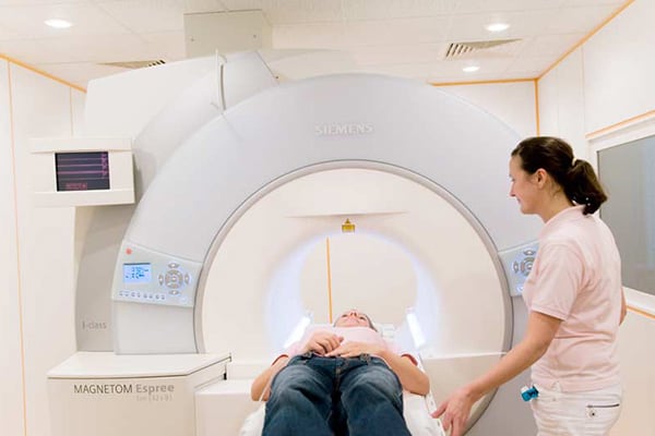
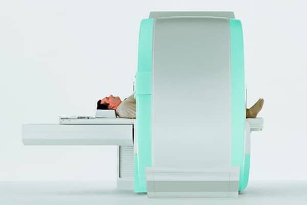
MR mammography – Breast cancer precaution with magnetic resonance imaging
The breast carcinoma is one of the most frequent malignant tumors of women in Germany (46.000 new cases per year). That implies a lifetime risk of 12 % for not handicapped women and 80 % for high-risk patients. 70 % of all carcinomas appear in patients without known risk factors. The tumor related rate of mortality is 40 %. Meanwhile more and more breast cancer specialists prefer the magnetic resonance imaging of the breast (so-called MR mammography), which can prove earlier and more reliable breast cancer focuses, than the classic conventional x-ray mammography. The MR mammography images the breast multidimensional, is without radiation and avoids the compression of the breast (which is necessary for x-ray and unpleasant for many women).
With the classical x-ray mammography, depending on the density of the breast tissue, only 20 to 80 % of all breast cancer results can be found, whereas the MR mammography detects 80 to 90 % of all cancer focuses ¬– regardless of the breast tissue structure. For comparison, the manual examination of the breast has a sensitivity of only 50 % and a minor specificity (accuracy of result detection). Self-examinations (own palpation) increase the biopsy rate, but not the sensitivity.
The classic x-ray mammography, has a remarkable bad sensitivity, especially for precursors of cancer (high grade ductal in situ carcinoma – DCIS), which do not get beyond 54 %, because a large part does not show microcalcifications. Altogether the sensitivity of the conventional mammography is only 28 to maximal 41 %, at a specificity of 98 %.
Therefore almost 60 % evade the diagnostics and from the 41 % detected tumors, 98 % are in average major carcinomas. The so-called interval carcinoma is usually indistinguishable in the preceded x-ray mammography. Frequent “false positive” results of the x-ray mammography can be reduced by the MR mammography: These are usually focuses with microcalcifications or overlays, which seem to be suspicious and therefore a biopsy is taken. In the end they are benign.
On the other side 30 to 40 % of all invasive carcinoma show microcalcifications. A secure classification if it is benign oder malignant is not possible with the x-ray mammography. Through inclusion of ultrasound the sensitivity increases to approximately 50 %. Whereas the MRI has a sensitivity of over 80 % and a specificity of approximately 90 %. It can detect tumors, which cannot be seen in the x-rays.
These numbers and further current studies show that the MR mammography not only detects breast cancer securer than the x-ray mammography, it can also detect it earlier. Even benign precursors of the invasive carcinoma can already be detected contrary to the x-ray mammography, which is primary only suitable to detect already existing invasive carcinomas.
The MR mammography leads to amendments of the treatment of already detected tumors, because in 18,75 % of the cases, additional contralateral tumors are found or the tumor spread is outlined different and in 8,7 to 9,7 % of the cases the surgical intervention is changed. Furthermore the MR mammography can gather premalignant atypical ductal hyperplasia with a sensitivity of 91 %, compared to 26 % of the conventional mammography.
Cost-benefit calculations already exist for high risk patients: When solely the conventional mammography is used, costs equivalent to 11.771 € per tumor arise. Costs of 9.024 € arise using MRI without x-ray.
The MR mammography can replace the so far used x-ray mammography in almost all cases. In circumstances where women have a pacemaker or other sensitive medical implants, the implementation of a MR mammography is not possible.


